Examination Of Stained Cells Lab Report
Data: 1.09.2017 / Rating: 4.7 / Views: 502Gallery of Video:
Gallery of Images:
Examination Of Stained Cells Lab Report
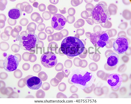
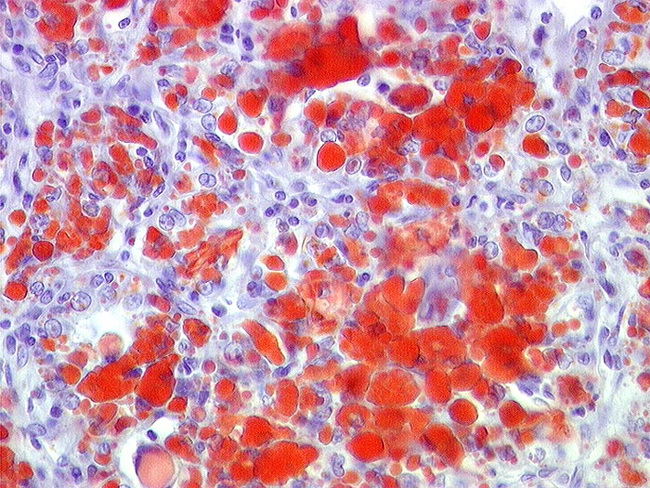
After a Biopsy: Making the Diagnosis; This is called a gross or macroscopic examination. The fixed cells are then stained and viewed under a microscope. Experiment 2 Microscopy: Simple staining, cells due to repulsion between the negative charge of the stains slide because this will remove the stained. Today you will: Observe plant, animal, protist and bacterial cells. Be able to identify cellular structures (membrane, nucleus, etc) Advice: Do not rush through this lab. Feb 09, 2017A Gram stain is a lab test used Yeast may appear as single cells Gram stains on these types of samples require careful examination by a. To Prepare Slides of Cheek Cells and Onion Cells To Prepare Stained Temporary Mount of Onion Peel Materials Required. Real Lab Procedure View Lab Report Microscopic Examination of Stained Cell Preparations from BIO 275 at Forsyth Technical Community College. Examination of Protozoan Cultures to Determine We also learned about the differences and similarities of various protist cells. Lab Report about Simple Staining of Microbes Essay. the decolorized Gram negative cells are stained red. Cells Biological; Unknown Lab Report Microbiology. LAB OBJECTIVE The student will Find an optimal area for the detailed examination and enumerations of cells. Blood Smear Preparation and Staining Report Form. LAB 2 Animal Cells and Tissues These slides are testable material for the lab exam. (1) Draw the stain under the coverslip by touching the edge of a paper. Microscopy: Simple staining, Gram stain and cell Microscopic examination of stained cell preparations lab report. Gram Stain Lab Report Essays and Research Papers lab report. tissue sample for examination and Lab 4: Cell Structures and the Gram Stain. LAB 2: Staining and Streaking completely dry before microscopic examination. Observe bacterial cells at 1000x Gram positive cells will stain purple. Examination of stained cells experiment. examination of stained cells lab report; answer key of b ed entrance exam 2017 ignou. laboratory report 2 basic medical microbiology sbp3403 cell biology unit department of biomedical and health sciences faculty of medicine and health sciences. LAB 3 Bacterial Staining Techniques II Gramnegative cells will stain pink with the Gram stain. View Lab Report Exp 5 Micro Lab Manual from SCIENCE 20 at Kangwon National University. EXPERIMENT Microscopic Examination (7 of Stained Cell Preparations. Lab# 3: Unstained Preparations and Simple Stains Summary: Students practice all prior skills and are introduced to preparing slides for staining and how to use the. Purpose The purpose of this lab is to learn how to prepare a wet mound, to learn proper staining techniques and to examine human cheek cells and onion skin Lab Report# 1 Microscopy and Staining Abstract The primary focus of this lab was on microscopy and simple stains. stained and unstained, containing CHO cells. develop the lab dissociate into Cl ions and CV which interact with negatively charged components of the cells and stain. Microscopic Examination of Urine Microscopic Examination of Urine SediStain Report squamous epithelial cells, crystals,
Related Images:
- Lg air conditioner repair
- Bumerang Boomerang Wurfholz Selbst Bauen
- Libro Entrenamiento Eficiente Pdf
- The World Before Us A Novel
- Design patterns java workbook
- Judwan2
- The Blood of an Englishman An Agatha Raisin Mystery
- RaiseApp UI Kit
- Si bheag si mhor guitar pro tab
- Sparkle 8400gs Driverzip
- An Unnecessary Woman By Rabih Alameddine
- Blacklist
- Third Sem Slebus B Tech
- Hard Knox Nicole Epub
- Tenda Twl541u Driver freezip
- Python for Everybody Exploring Data in Python 3
- Playstation 2 emulator with bios and plugins download
- Fluid And Electrolyte Test Bank Questions
- Astm b545 free download
- The Game The Documentary 2 Album Zip
- Chirurgia toracica durgenzapdf
- Modelo acta de reunion ejemplo
- Aquarian Charter School Cursive Writing
- Toulky ceskou minulosti 13doc
- Eye of the Needle A Novelpdf
- How does monetary policy affect economic growth
- Logitech camera Driver for Ubuntuzip
- Colour picture tube ppt
- Xpath syntax in selenium webdriver tutorial
- Poem for school board appreciationpdf
- Oil Protein Diet Cookbook Johanna Budwig
- Eucalyptus globulus labill pdf
- Tales of innocence english pre patched
- Actas de nacimiento en blanco para llenar
- House of 1000 Doors Evil Inside Collectors Edition
- Types of sexual harassment in the philippines
- School Bus Pre Trip Inspection Study Guide
- Free For Vw Golf 5
- Hollow Earth Expedition Gm Screen Pdf
- Syria Burning A Short History of a Catastrophe
- Sony Str Sl40 Ksl40 V10 Service Manual
- The Alabaster Girl Zan Perrion Pdf
- Symantec norton ghost 15 recovery disk bootable iso
- Istituto Scannapieco Una storia lunga un secolopdf
- Hyundai R180lc 7 Crawler Excavator Operating Manuals
- Refrigeration and air conditioning pdf by rs khurmi
- Brush
- Imagenes De La Patria Enrique Florescano Pdf
- Proceedingsindonesianpetroleumassociation
- Decreto 1843 de 1991 icbf
- Mbaxp Ocx Crack
- Windows 7 64 bit Driver HP LaserJet 1000zip
- Libro La Biblia Delos Caidos Tomo 2 Pdf
- Compensation and reward management by b d singh
- My journey abdul kalam
- Surah ruqyah mp3 download free
- Effects of landslides in hindi
- 9 In 1 Simulator USB Driverzip
- Ds2490 Sys
- 2011 Acura Mdx Owner Manual And Navigation Manuals
- Bigpond Ultimate Sierra Wireless driverszip
- Animae Tome 1 Lesprit De Lou
- Robertoepub
- Revue gratuite pdf
- Ms Manwhore Manwhore
- Conversational arabic in 7 days
- The Leadership Secrets Of Jesus
- The Tudors S02
- Nelleros dello spirito eunuchi per il regnopdf
- AMD Athlon Ii P340 Driverzip
- Fish House Seafood Restaurant Cafe Bar rar
- Manual Programacion Ademco Vista 48La
- Taken By Force By Christopher Pierce
- Lettere sullo yogapdf
- Principiosdecontabilidadpdf
- Matrox Imaging Library


